Humanness Analysis
Evaluates antibody humanness using OASign (Observed Antibody Space Immunogenicity) and PFA (Positional Frequency Analysis) to quantify how "human-like" a sequence is based on natural antibody repertoires.
[!IMPORTANT] Integrated Immunogenicity Assessment: Sapiens, OASign, and RPEMHC must be evaluated in conjunction for a complete developability profile. OASign and Sapiens quantify sequence humanness (which corresponds to T-cell receptor self-tolerance), whereas RPEMHC predicts physical peptide presentation to human MHC-II. A therapeutic candidate is only safe if it scores well on both metrics: a "Good" (Green) RPEMHC score on a sequence with low humanness (such as a mouse sequence) is still highly immunogenic in patients because there is no self-tolerance for the presented peptides. Furthermore, predicted antigen-binding contacts from Paragraph are integrated across all diagnostic cards, tables, and alignments to safeguard target binding affinity during de-immunization engineering.
Humanness Analysis functions as a De-Immunization and Engineering Mutation Guide. Rather than attempting to serve as a continuous predictor of clinical trial Anti-Drug Antibody (ADA) percentages (which depend on patient immunological status, disease indication, concomitant medications, subcutaneous vs. intravenous administration, and sub-visible aggregate particles), this tool isolates the specific HLA Class II presentation hotspots, non-human 9-mer stretches, and sequence outliers driving immunogenic risk across the variable domain (\(F_v\)). When an antibody exhibits immunogenicity liabilities or requires humanization, engineers use this workspace to pinpoint specific residues to substitute to abrogate MHC-II groove anchoring or restore human repertoire consensus while preserving antigen binding.
Why Humanness Functions as a Mutation Guide Rather Than an Assay Predictor
In silico immunogenicity models cannot reliably predict continuous numerical rates of clinical anti-drug antibody (ADA) development:
-
Clinical and Patient Diversity: Clinical ADA development is an emergent immunological outcome shaped by the patient's immune competence (e.g., immunosuppressed oncology patients vs. hyperactive autoimmune patients), background genetics (diverse HLA allotypes), and concomitant therapies. For example, co-administration of methotrexate reduces ADA incidence against anti-TNF antibodies from \(>30\%\) to \(<5\%\) without altering the antibody sequence.
-
Route of Administration and Regimen: Subcutaneous (SC) injection delivers therapeutic proteins into tissue beds dense with professional antigen-presenting cells (dendritic cells) and lymphatic drainage, resulting in significantly higher ADA incidence than intravenous (IV) infusion for identical antibody molecules. Repeated chronic administration also drives higher rates of secondary ADA responses than single-dose regimens.
-
Formulation and Sub-Visible Particles: Sub-visible particles (\(1\text{--}10\ \mu\text{m}\)) and soluble aggregate species act as potent self-adjuvants. By cross-linking B-cell receptors (BCR), aggregates can elicit robust ADA responses even against \(100\%\) germline human antibodies.
-
Central and Peripheral Tolerance: Neural network MHC-II binding predictors (such as RPEMHC) evaluate the physical chemical affinity of peptides into polymorphic Class II grooves, but they cannot model thymic negative selection (central clonal deletion), regulatory T-cell networks (Tregitopes), or anergy.
Because continuous numerical regressions of clinical ADA percentages provide false precision, AbLead focuses on epitope hazard identification: detecting the physical 15-mer peptide anchors and sequence liabilities on the variable domain so engineers know exactly which residues to substitute to remediate immunogenicity liabilities while safeguarding antigen binding affinity.
Accessing the Tool
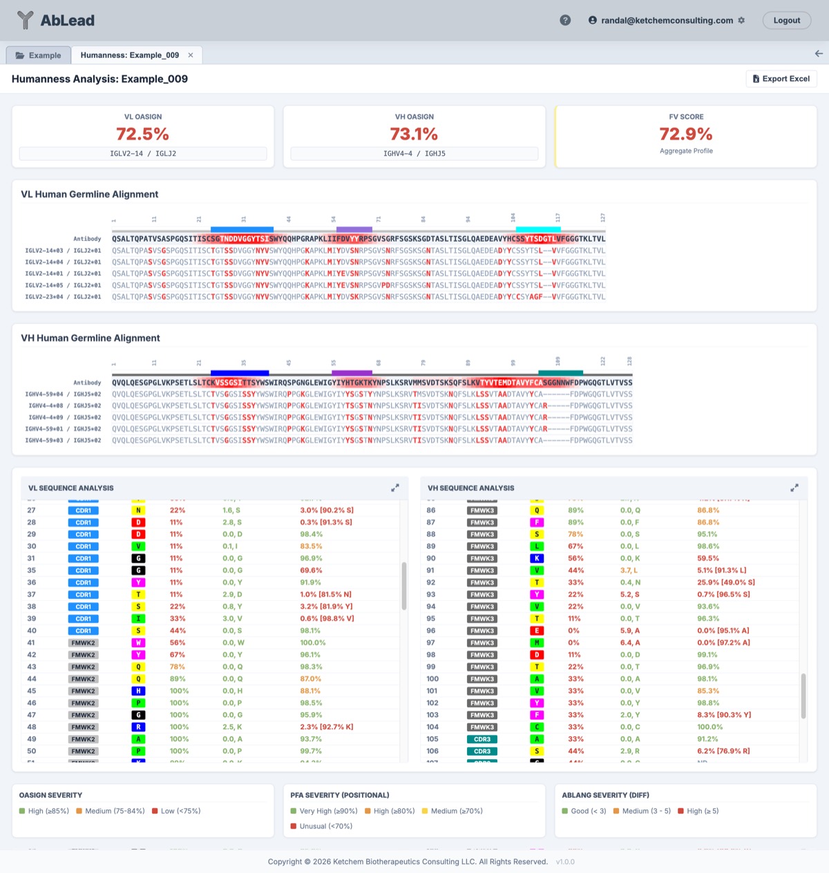
Select an antibody in the Project View. Go to the Analysis menu and select Humanness. This will open the Humanness Analysis workspace.
Click the Mutation Guide button in the top toolbar to view a reference modal detailing triage thresholds, anchor-breaking substitution strategies, the 4 quadrants of immunogenicity risk, and paratope preservation tips.
Using the Tool
The Humanness Analysis workspace provides an integrated, side-by-side view combining OASign repertoire sequence humanness with RPEMHC Class II MHC presentation risk alongside granular residue-level diagnostics.
1. Unified Aggregate Scoring (Two Rows)
-
Row 1 — OASign Repertoire Humanness:
- VL / VH OASign (%): The fraction of 9-mer peptides in the variable region that appear in natural human antibody repertoires.
- Fv OASign (%): The peptide-weighted aggregate humanness of the paired variable domain.
- Germline Gene Boxes: Automatically displays the closest matching human V and J germlines.
-
Row 2 — RPEMHC ADA Immunogenicity Risk:
- VL / VH RPEMHC: The 4-decimal probability (\(0.0000 – 1.0000\)) that the chain variable domain triggers helper T-cell activation.
- Fv RPEMHC: The paired Fv anti-drug antibody (ADA) probability, color-coded by clinical risk (\(\le 0.35\) Green, \(\le 0.45\) Orange, \(> 0.45\) Red).
- Allele Context: Displays the top driving HLA Class II allotype for the active panel.
2. High-Risk MHC-II Epitopes (T-Cell Hotspots)
- Hotspot Extraction and 1-Click Noise Filter: Detects high-affinity 15-mer sliding windows (\(\ge 0.638\)) across the Fv domain.
- Human Germline Context: Badges each epitope as
100% Germline(tolerated self-peptide),Somatic/Point Mutation,CDR3 Junction, orNon-Germline. - Predicted Paratope Overlap: Highlights hotspots overlapping predicted antigen-binding contacts (\(\ge 50\%\) probability via Paragraph) with a blue
Pbadge in the Region column. The header displays a summary badge (X Paratope) indicating the total count of paratope-overlapping hotspots. - 1-Click Noise Reduction and Paratope Filters: Use filter buttons to toggle between All Hotspots, Non-Germline Only (eliminating noise to focus strictly on actionable de-immunization liabilities), Germline Only, and Paratope (isolating epitopes containing critical paratope residues to avoid reducing target affinity during de-immunization).
- Interactive Multi-Column Sorting: Click any column header (Chain, Region, Positions, Peptide, Germline Status, Top Allele, Peak Binding Score, Population Breadth) to cycle Ascending \(\rightarrow\) Descending \(\rightarrow\) Remove. Supports composite multi-column sorting with precedence badges (
[1],[2], etc.) and default binding score ranking.
3. Dual-Track Sequence Alignment Cards
- Region Bar Track: Displays structural region assignments (framework and CDR loops) across light and heavy chains.
-
Predicted Paratope Track: A dedicated 10px strip situated directly below the Region Bar row, color-coded by Paragraph contact probability:
- Contact (\(\ge 50\%\)): Deep vibrant blue (
#1d4ed8), indicating high-confidence predicted paratope contact residues. - Intermediate (\(20\%–49\%\)): Soft blue (
#93c5fd), representing moderate contact probability or paratope periphery. - Low / Non-contact (\(< 20\%\)): Neutral light gray (
#e2e8f0), indicating residues with negligible contact likelihood. - Interactive Tooltips and Scrolling: Hovering displays the position, residue, contact probability percentage, and confidence level. Clicking any cell immediately scrolls the corresponding Sequence Analysis table to that residue with a flash outline highlight.
- Contact (\(\ge 50\%\)): Deep vibrant blue (
-
Query (OASign) Track: Colored by non-human 9-mer clusters (white for 100% human, yellow \(\rightarrow\) red for non-human stretches). Hovering over any residue displays overlapping 9-mers and mutations required to reach human sequences.
- Query (RPEMHC) Track: Colored by MHC-II groove binding affinity for the active allele panel (\(\ge 0.638\) orange, \(\ge 0.80\) red).
- Matched Germline Tracks: Displays aligned human germline reference sequences with non-germline mutations highlighted in red.
4. Residue-Level Analysis (Dual-Pane)
The diagnostic tables in the VL and VH panes display both metrics simultaneously:
- Region and Predicted Paratope (
Reg): Displays framework and CDR loop assignments alongside a bold bluePbadge for residues identified as predicted antigen-binding contacts (\(\ge 50\%\) probability via Paragraph). Hovering displays the contact probability tooltip warning of potential binding impact. - OASign (%): Percentage of human 9-mers overlapping this residue.
- MHC-II Score: Maximum predicted Class II binding score overlapping this residue, with tooltips showing the top presenting HLA allele.
- Sapiens Diff: Difference between actual residue probability and highest-scoring human alternative predicted by the Sapiens model.
- PFA: Positional frequency analysis against human germlines (with hover tooltip displaying the full metric description).
- Engineering Modal: Clicking any residue opens the Engineering modal with predictive de-immunization scans and live score previews. The scan table evaluates all 20 amino acid substitutions across predicted MHC-II binding, human germline frequency (PFA), and human repertoire probability (Sapiens).
- Unified Excel Export: One-click export generates a comprehensive workbook with summary cards, dual OASign/RPEMHC columns, 20 allele scores, and an Fv Hotspots sheet.
Strategy: Triage positions using the Heatmap. Use PFA and Sapiens Diff scores to find weak spots. Open the Engineering modal on high-risk residues to inspect the predictive scans and select human-favored substitutions that lower predicted MHC-II binding scores without disrupting structural properties.
Scoring Thresholds
AbLead uses standardized clinical thresholds to categorize OASign and RPEMHC results:
| OASign Score | Category | UI Color (colors.py) |
Description |
|---|---|---|---|
| 85% – 100% | High / Good | Green (#88B46C) |
Matches the mean score of natural human germline antibodies. |
| 75% – 84% | Medium / Low Risk | Yellow (#F8D548) |
Typical range for clinically successful humanized antibodies. |
| < 75% | Low / High Risk | Red (#CF4A3C) |
Below standard clinical humanization norms. |
| RPEMHC Probability (Single Chain / Fv) | Category | UI Color (colors.py) |
Description |
|---|---|---|---|
RPEMHC VL / VH \(\le 0.2000\) (Fv \(\le 0.3500\)) |
Good / Low Risk | Green (#88B46C) |
Low population-weighted helper T-cell activation probability. |
RPEMHC VL / VH \(0.2000–0.3000\) (Fv \(0.3500–0.4500\)) |
Warning / Moderate | Yellow (#F8D548) |
Moderate clinical risk range / candidate T-cell neo-epitopes. |
RPEMHC VL / VH \(> 0.3000\) (Fv \(> 0.4500\)) |
Severe / High Risk | Red (#CF4A3C) |
High helper T-cell activation probability / elevated ADA risk (flags Bococizumab at 0.4630 in RED). |
| PFA Score | Category | UI Color | Description |
|---|---|---|---|
| ≥ 90% | Very High | Green | Highly conserved human germline residue. |
| ≥ 80% | High | Orange | Common human germline residue. |
| ≥ 70% | Medium | Yellow | Moderately common human germline residue. |
| < 70% | Unusual | Red | Infrequent or unusual residue at this position. |
| Sapiens Diff | Category | UI Color | Description |
|---|---|---|---|
| < 0.15 | Good | Green | High human consensus; residue matches or is close to top human alternative. |
| 0.15 – 0.35 | Medium | Orange | Moderate divergence from human antibody repertoires. |
| ≥ 0.35 | High | Red | Significant divergence from human repertoires; primary target for humanization. |
| Sapiens Scan Probability | Category | UI Color | Description |
|---|---|---|---|
| ≥ 50% | Favored / High | Green (#10b981) |
Frequently observed substitution in natural human antibody repertoires. |
| 10% – 49% | Moderate | Yellow / Orange (#eab308) |
Permissible human repertoire substitution. |
| < 10% | Rare / Disfavored | Red (#ef4444) |
Rare or disfavored in human repertoires; potentially non-human or destabilizing. |
| Predicted Paratope Probability | Category | UI Color (colors.py) |
Description |
|---|---|---|---|
| ≥ 50% | Contact | Deep Blue (#1d4ed8) |
High probability antigen-binding paratope contact (P badge). |
| 20% – 49% | Intermediate | Soft Blue (#93c5fd) |
Moderate contact likelihood or paratope periphery. |
| < 20% | Low / Non-contact | Neutral Gray (#e2e8f0) |
Negligible predicted binding contact. |
Scientific and Empirical Basis for Thresholds
- OASign Thresholds (\(\ge 0.8500\), \(0.7500–0.8400\), \(< 0.7500\)): Derived from the BioPhi / OASis benchmark study (Prihoda et al., mAbs 2022). Natural human antibodies average \(\ge 85\%\) repertoire identity. Clinically approved humanized antibodies (e.g. Trastuzumab, Bevacizumab, Pertuzumab) score between \(75\%\) and \(85\%\), while non-human/murine sequences fall below \(75\%\).
- RPEMHC Population Probability Thresholds (VL/VH \(\le 0.2000\), Fv \(\le 0.3500\)): Calibrated using first-principles biophysical groove binding activation and global HLA Class II human population genetics benchmarks (Paul et al., Immunity 2013). Clinically approved low-ADA therapeutics display clean profiles (\(\le 0.3500\) Fv / \(\le 0.2000\) VL/VH), while high-risk candidates (e.g. Bococizumab at \(0.4630\) Fv) are flagged with severe developability penalties.
- PFA Conserved Residue Cutoffs (\(\ge 90\%\), \(\ge 80\%\), \(\ge 70\%\), \(< 70\%\)): Based on IMGT human germline amino acid distributions across matched V and J gene families. Residues \(<70\%\) represent non-canonical framework variations, murine backmutations, or somatic mutations.
- Sapiens Human Likelihood Difference (\(\Delta < 0.15\), \(0.15–0.35\), \(\ge 0.35\)): Evaluated using the Sapiens human antibody language model (Prihoda et al., mAbs 2022), where \(\Delta = P_{\text{top human}} - P_{\text{actual}}\) flags non-human positions and provides direct parity with mutations recommended in the Humanization tool.
- Paragraph Paratope Contact Cutoffs (\(\ge 50\%\), \(20\%–49\%\), \(< 20\%\)): Derived from the Paragraph equivariant graph neural network benchmark (Deszyński et al., Bioinformatics 2023). Residues scoring \(\ge 50\%\) represent high-confidence antigen-binding contacts, while \(20\%–49\%\) captures peripheral or permissive contact residues. Protecting \(\ge 50\%\) residues during de-immunization engineering prevents accidental loss of antigen binding affinity and functional potency.
The 4 Quadrants of Immunogenicity Risk
Clinical immunogenicity (Anti-Drug Antibody / ADA development) is governed by two orthogonal biological components:
- Biophysical Groove Presentation (RPEMHC): Does the peptide anchor into human HLA Class II pockets with sufficient affinity to be displayed by antigen-presenting cells?
- Repertoire Tolerance vs. Foreignness (OASign / PFA): Is the presented peptide recognized as natural human "Self" (tolerized during thymic negative selection) or "Foreign" (stimulating naive CD4+ T-cells)?
| High OASign (\(\ge 0.8500\) Human Repertoire) | Low / Moderate OASign (\(< 0.8000\) Non-Human / Foreign) | |
|---|---|---|
| Low RPEMHC (\(\le 0.3500\)) | Ideal Clinical Profile (e.g., Trastuzumab, Bevacizumab) • Fully tolerized by human immune system • Low Class II presentation density • Lowest statistical risk of clinical ADA |
De-Immunized Non-Human / Chimeric • Sequence is foreign/non-human • Lacks physical MHC-II groove anchors • Minimal presentation to helper T-cells |
| High RPEMHC (\(> 0.4500\)) | Self-Presenter (e.g., Renvistobart) • High biophysical groove affinity • Composed of natural human repertoire 9-mers • Tolerized by central/peripheral immune mechanisms |
Clinical Danger Zone (e.g., Bococizumab) • Strong MHC-II peptide presentation (\(0.4630\), Red) • Contains non-human / foreign CDR neo-epitopes • High risk of helper T-cell priming & neutralizing ADAs |
Clinical Case Study: Bococizumab (The Danger of Global Score Averaging)
Bococizumab (Pfizer’s humanized anti-PCSK9 mAb) provides a classic clinical example of why granular epitope evaluation is essential:
- Clinical Outcome: In late-stage Phase 3 trials (the SPIRE program, encompassing over 27,000 patients), Pfizer was forced to discontinue development after discovering that 48% of patients developed anti-drug antibodies (ADAs), with 29% developing high-titer neutralizing ADAs that substantially eroded LDL-C lowering efficacy over time and caused elevated injection-site reactions.
-
First-Principles Immunogenic Profile: Under AbLead's population-weighted probability model, Bococizumab is unequivocally flagged as a high-risk candidate:
OASign Fv: \(0.8134\) (standard humanized range; yellow)RPEMHC VL: \(0.2540\) (warning / moderate; yellow)RPEMHC VH: \(0.2360\) (warning / moderate; yellow)RPEMHC Fv: \(0.4630\) (severe / high risk; red)
-
The Immunological Mechanism: The human immune system does not evaluate global sequence averages. Bococizumab’s CDR loops (derived from a murine precursor) contained non-human somatic mutations that formed high-affinity 15-mer peptide anchors (\(\ge 0.800\)) presented across multiple core HLA-DRB1 alleles. Because these foreign CDR peptides had never undergone thymic negative selection in humans, dendritic cells presented them to naive CD4+ T cells, priming T follicular helper (\(T_{\text{FH}}\)) cells and driving robust B-cell class switching to neutralizing anti-bococizumab IgG antibodies.
- Key Engineering Takeaway: Global Fv scores provide high-throughput triage, but lead candidates must always be evaluated at the residue and sliding-window level using the RPEMHC 2D Matrix Heatmap, Linear Sequence Track, and Engineering Modal to detect and eliminate isolated CDR helper T-cell hotspots prior to clinical commitment.
High-Risk MHC-II Epitopes (Hotspots Card)
To prevent localized immunogenic liabilities from being masked by whole-chain averages, the Humanness workspace in RPEMHC Mode includes a dedicated High-Risk MHC-II Epitopes (Hotspots) summary table:
- Detection Criteria: Automatically extracts 15-mer peptide windows exhibiting high groove binding affinity (\(\ge 0.638\)) or broad population promiscuity (\(\ge 4\) presenting HLA Class II alleles).
- Human Germline Context and Self-Tolerance: Distinguishes 100% human germline peptides (which are naturally tolerized by central immune tolerance) from true neo-epitope liabilities containing somatic hypermutations (SHM), point mutations, or CDR3 junctions.
- Germline Status Badges and Tooltips: Displays clear visual origin badges (
100% Germline,1 Mut: Pos,CDR3 Junction, orNon-Germline). Hovering over any badge or peptide provides an interactive tooltip showing the query sequence vs. matched human germline sequence, assigned V/J gene family, and exact amino acid mutations. - Predicted Paratope Contact Identification: 15-mer epitopes that overlap one or more predicted antigen-binding paratope residues (\(\ge 50\%\) probability via Paragraph) are tagged with a bold blue
Pbadge in the Region column. The card header shows a summary badge (X Paratope) with the total number of paratope-overlapping hotspots. -
1-Click Filtering Controls: Allows users to filter the table instantly between:
- All Hotspots: Displays all identified high-affinity epitopes across both chains.
- Non-Germline Only: Focuses de-immunization engineering purely on actionable, non-tolerized neo-epitopes.
- Germline Only: Isolates tolerized native germline peptides.
- Paratope: Filters strictly for epitopes that overlap predicted antigen-binding paratope residues, highlighting liabilities that require careful de-immunization to avoid disrupting antigen binding.
-
Clustered Peak Reporting: Groups contiguous overlapping 15-mers into distinct physical epitope clusters, reporting the peak 15-mer peptide sequence, top presenting HLA allotype, peak binding score (color-coded by risk severity), and total population breadth (out of 20 alleles).
- Interactive Triage: Clicking any hotspot row immediately scrolls both the sequence alignment and residue table to that exact position and applies an outline highlight to facilitate immediate de-immunization engineering.
Positional Max Risk Evaluation
When viewing sequence alignments and residue diagnostics under Max Risk — Tier 1 Global Panel or Max Risk — All 20 Alleles:
- Positional Maximum Evaluation: Rather than fixing the entire sequence to the single allele that had the highest overall chain total sum, each individual amino acid position evaluates the true maximum binding score across all panel alleles at that specific position.
- Allele Attribution Tooltips: Hovering over any residue box in the alignment or the MHC-II score cell in the table displays the exact HLA Class II allotype driving the peak risk (e.g.
MHC-II Score (Max Tier 1: DRB1*04:05): 1.0030), immediately revealing localized neo-epitopes.
Methodology
The AbLead Humanness analysis suite combines repertoire database lookups, germline positional frequencies, masked language models, and deep neural network Class II MHC binding predictors to create an integrated developability and immunogenicity assessment.
1. OASign: Repertoire-Based Sequence Humanness
OASign measures sequence humanness by comparing antibody peptide fragments directly against the Observed Antibody Space (OAS), a comprehensive repository of over one billion natural human antibody sequences derived from diverse healthy donors.
- Fv Domain Trimming: Sequences are automatically identified and trimmed to the Variable Domain (\(\text{Fv}\)) using AntPack annotation. Constant domains are excluded from repertoire scoring to prevent false inflation from invariant constant sequences.
- Sliding 9-mer Peptide Decomposition: The variable region sequence of length \(L\) is chopped into \(L - 8\) overlapping 9-mer peptide windows. A 9-mer window size is biologically calibrated to match the typical footprint of linear B-cell epitopes and linear sequence identity segments.
- Hash-Based Repertoire Querying: Each 9-mer is queried against a high-performance hash index of human antibody repertoires. A 9-mer is scored as Human (1.0) if it appears in the curated human OAS database with sufficient donor support, and Non-Human (0.0) otherwise.
- Mutational Distance to Human: For any non-human 9-mer, OASign computes the minimum single amino acid substitution required to convert the peptide into a known human repertoire 9-mer, identifying minimal-mutation humanization pathways.
-
Chain & Fv Humanness Scores: The percentage of human 9-mers in the variable domain:
\[\text{Score}_{\text{Chain}} = \frac{\sum_{i=1}^{N_{\text{peptides}}} I(\text{peptide}_i \in \text{OAS})}{N_{\text{peptides}}} \times 100\%\]\[\text{Score}_{\text{Fv}} = \frac{N_{\text{pep, VL}} \cdot \text{Score}_{\text{VL}} + N_{\text{pep, VH}} \cdot \text{Score}_{\text{VH}}}{N_{\text{pep, VL}} + N_{\text{pep, VH}}}\]
2. Positional Frequency Analysis (PFA)
Positional Frequency Analysis evaluates how standard or unusual an amino acid is at a specific topological position compared to natural human antibody germlines.
- IMGT Alignment: Sequences are aligned against IMGT human germline reference databases for heavy (
IGHV,IGHJ) and light (IGKV,IGLV,IGKJ,IGLJ) chains. - Positional Matrix Lookups: For each aligned IMGT position, the system looks up the frequency distribution of all 20 amino acids across human germlines belonging to the matching V and J gene families.
- Frequency Output: Reports the percentage of human germline antibodies carrying the query residue alongside the dominant human amino acid at that position (e.g.
0.5% [90.7% T]). Positions with low PFA scores (\(<70\%\)) represent somatic hypermutations, non-canonical framework substitutions, or murine backmutations that may destabilize the antibody or act as foreign epitopes.
3. Sapiens Human Antibody Language Model
Sapiens is a deep learning masked language model trained strictly on tens of millions of human antibody sequences from the Observed Antibody Space (OAS) (Prihoda et al., mAbs 2022). Unlike species-agnostic antibody language models (which can score murine sequences as normal), Sapiens models the specific sequence manifold of human antibodies.
-
Human Contextual Probability: Given the full variable domain, Sapiens predicts the human repertoire probability distribution across all 20 amino acids at each position (\(P_{\text{actual}}\)).
-
Difference Metric (\(\Delta = P_{\text{top human}} - P_{\text{actual}}\)): Evaluates the difference between the optimal human residue probability and the actual residue probability:
\[\Delta = P_{\text{top human}} - P_{\text{actual}}\]A low difference (\(\Delta < 0.15\)) indicates that the residue is favored by human repertoire consensus, whereas a high difference (\(\Delta \ge 0.35\)) flags positions that diverge significantly from human antibodies and represent prime candidates for humanization.
-
Predictive De-Immunization Scanning: In the residue engineering modal, Sapiens repertoire probability percentages (\(0.0\% - 100.0\%\)) are computed for all 20 amino acid substitutions, color-coded by human repertoire frequency (\(\ge 50\%\) Green, \(10\% - 49\%\) Yellow/Orange, \(< 10\%\) Red). This enables protein engineers to identify de-immunizing substitutions that reduce Class II MHC groove presentation while ensuring high human repertoire compatibility.
4. RPEMHC: Relative Position Encoding for MHC-II Binding
RPEMHC predicts the physical chemical presentation of 15-mer antibody peptides to human Major Histocompatibility Complex Class II (MHC-II) molecules.
- 15-mer Sliding Windows: Antibody sequences are scanned using a sliding window of 15 amino acids (\(L - 14\) windows per chain), capturing the full peptide length that extends beyond the open-ended MHC-II binding groove.
- Relative Position Encoding: Unlike standard sequence encoding, relative position encoding explicitly represents the spatial distance between amino acid pairs and the polymorphic MHC-II binding pockets (P1, P4, P6, P7, P9), capturing cooperative anchoring interactions.
- 20-Allele Panel Evaluation: Evaluates binding affinity across both Tier 1 Core DRB1 alleles (>95% global population coverage) and Tier 2 Extended alleles (DRB3/4/5, DQ, DP).
- Thresholding and Cumulative Epitope Load: Peptides scoring \(> 0.50\) are flagged as presenting helper T-cell epitopes. The per-allele and aggregate construct scores (\(\sum \text{Scores} > 0.50\)) quantify total immunogenic burden to guide de-immunization engineering.
5. Paragraph: Deep Learning Paratope Prediction
Paratope prediction identifies the residues on an antibody variable domain that directly engage the target antigen. In AbLead, paratope predictions are powered by Paragraph, an Equivariant Graph Neural Network (EGNN) developed by the Oxford Protein Informatics Group (OPIG) (Deszyński et al., Bioinformatics 2023).
- Structure-Aware Graph Neural Network: Paragraph models the 3D atomic coordinates of antibody variable domains (Fv), analyzing spatial geometry, solvent accessibility, and CDR surface conformations to predict antigen-binding likelihood without requiring prior knowledge of the target epitope.
- Residue-Level Contact Probabilities: Outputs continuous probability values between \(0.000\) and \(1.000\) for each residue in the variable domain. Residues with probabilities \(\ge 0.500\) are flagged as core paratope contacts.
- Guiding De-Immunization Engineering: Pairing RPEMHC T-cell hotspot predictions with Paragraph paratope contacts allows protein engineers to de-risk de-immunization campaigns. Hotspots situated entirely within framework regions or non-contact CDR loops can be mutated safely toward human germline consensus, whereas hotspots overlapping predicted paratope residues (
Pbadge) require targeted substitutions that preserve critical antigen-binding interactions.
Understanding HLA Class II Alleles and Nomenclature
1. WHO / IMGT-HLA Nomenclature (e.g., HLA-DRB1*04:01)
Allele designations follow the standard World Health Organization (WHO) and IMGT-HLA nomenclature:
- Gene Locus: Prefix (e.g.,
HLA-DRB1*,HLA-DQA1*,HLA-DPA1*) designates the Class II \(\alpha\) or \(\beta\) chain gene. - Field 1 (e.g.,
04): The broad serological antigen group / allele family (e.g., DR4). - Field 2 (e.g.,
:01,:05): The specific protein allotype / subtype. Specific amino acid polymorphisms between subtypes alter the chemical topology and charge distribution of peptide-binding pockets (P1, P4, P6, P7, P9), resulting in distinct epitope repertoires.
2. Tier 1 vs. Tier 2 Allele Panels
- Tier 1 (Core Global Reference Panel): 11 high-prevalence
HLA-DRB1alleles (01:01,03:01,04:01,04:05,07:01,08:02,09:01,11:01,12:01,13:02,15:01) representing over 95% cumulative coverage across diverse global human populations. - Tier 2 (Extended Panel): 9 additional high-impact alleles across secondary
DRB3/4/5,DQA1/DQB1, andDPA1/DPB1loci for complete HLA Class II immunogenicity risk profiling.
3. HLA Class II Reference Panel
| Allele (WHO / IMGT) | Panel / Tier | Estimated Population Frequency | Binding Pocket Motif Preferences | Clinical / Therapeutic Risk Associations |
|---|---|---|---|---|
| HLA-DRB1*01:01 | Tier 1 Core | ~10–15% Global (EUR ~15–20%, ASN ~5–10%) | P1: Large hydrophobic (F, Y, W, L, I); P4/P6/P9: Aliphatic | Broad immunogenic peptide presentation; common ADA epitope presenter |
| HLA-DRB1*03:01 | Tier 1 Core | ~10–15% Global (EUR ~12–18%, AFR ~10–15%) | P1: Hydrophobic (L, I, M, F); P4: Acidic (D, E); P6: Basic/polar | Associated with SLE, Type 1 diabetes, autoimmune thyroiditis, biological drug ADA |
| HLA-DRB1*04:01 | Tier 1 Core | ~8–14% Global (EUR ~15–20%, AMR ~10–15%) | P1: Aromatic/aliphatic (F, Y, W, L); P4: Basic/polar; P9: Hydrophobic | Shared epitope for Rheumatoid Arthritis; high clinical ADA risk in TNFα biologics |
| HLA-DRB1*04:05 | Tier 1 Core | ~5–10% Global (East Asian ~15–25%, EUR ~2–4%) | P1: Aromatic/aliphatic; P4: Acidic/polar (pocket 4 subtype difference) | Key Asian-predominant RA & therapeutic immunogenicity risk allele |
| HLA-DRB1*07:01 | Tier 1 Core | ~15–22% Global (EUR ~25%, Middle Eastern ~25%) | P1: Large aromatic (F, Y, W); P4: Large hydrophobic; P9: Aliphatic | High frequency globally; co-expressed with DRB4*01:01; frequent mAb ADA epitope |
| HLA-DRB1*08:02 | Tier 1 Core | ~3–8% Global (Hispanic/Amerindian ~10–20%, ASN ~5–10%) | P1: Aliphatic/aromatic; P4: Acidic/polar; P6: Polar; P9: Hydrophobic | Important representation for Hispanic/Latin American and Indigenous cohorts |
| HLA-DRB1*09:01 | Tier 1 Core | ~5–10% Global (East Asian ~15–20%, EUR ~2–5%) | P1: Large hydrophobic; P4: Polar/aliphatic; P9: Small neutral | Prominent in East Asian populations; associated with autoimmune hepatitis & ADA |
| HLA-DRB1*11:01 | Tier 1 Core | ~10–18% Global (Mediterranean ~20–25%, AFR ~10–15%) | P1: Aliphatic/aromatic; P4: Small/polar; P6: Basic/polar; P9: Aliphatic | High Mediterranean and Hispanic frequency; co-expressed with DRB3*02:02 |
| HLA-DRB1*12:01 | Tier 1 Core | ~3–8% Global (East Asian ~8–15%, AFR ~5–10%) | P1: Hydrophobic; P4: Polar/basic; P9: Aliphatic | Asian and African cohort diversity representation |
| HLA-DRB1*13:02 | Tier 1 Core | ~6–12% Global (AFR ~8–15%, EUR ~6–10%) | P1: Hydrophobic; P4: Acidic/polar; P6: Polar; P9: Aliphatic | Broad peptide binding capacity; protective in certain viral/autoimmune contexts |
| HLA-DRB1*15:01 | Tier 1 Core | ~12–20% Global (EUR ~20%, ASN ~15–20%) | P1: Hydrophobic/aromatic; P4: Aliphatic; P7: Polar; P9: Small neutral | Strong association with Multiple Sclerosis; major driver of therapeutic protein ADA |
| HLA-DRB3*01:01 | Tier 2 (DRB3) | ~15–30% (Haplotype-linked with DR3, DR11, DR13, DR14) | P1: Hydrophobic; P4: Basic; P6/P9: Aliphatic | Co-expressed secondary β-chain; contributes additive Class II presentation |
| HLA-DRB3*02:02 | Tier 2 (DRB3) | ~20–35% (Co-expressed with DR11, DR13, DR14) | P1: Aliphatic/aromatic; P4: Polar; P9: Aliphatic | High expression in individuals carrying DRB1*11:01 / DRB1*13:01 haplotypes |
| HLA-DRB4*01:01 | Tier 2 (DRB4) | ~30–45% (Haplotype-linked with DR4, DR7, DR9) | P1: Hydrophobic; P4: Neutral/aliphatic; P7/P9: Polar | Highly prevalent secondary DRB molecule in DR4/DR7 carriers; prominent ADA mediator |
| HLA-DRB5*01:01 | Tier 2 (DRB5) | ~15–25% (Haplotype-linked with DR15, DR16) | P1: Large aromatic; P4: Polar/aliphatic; P9: Hydrophobic | Co-expressed with DRB1*15:01; implicated in MS and myelin peptide presentation |
| HLA-DQA1*05:01 / DQB1*02:01 | Tier 2 (DQ2.5) | ~10–20% Global (EUR ~20%, Hispanic ~15%) | P1/P9: Hydrophobic; P4/P6/P7: Negative charge (Glu, Asp, deamidated Gln) | Canonical DQ2.5 heterodimer; strong celiac disease & deamidated therapeutic epitope risk |
| HLA-DQA1*03:01 / DQB1*03:02 | Tier 2 (DQ8) | ~10–18% Global (AMR ~20–30%, EUR ~15%) | P1/P9: Hydrophobic/acidic; P4: Negative/polar | Canonical DQ8 heterodimer; associated with autoimmune diabetes and specific ADA |
| HLA-DQA1*01:02 / DQB1*06:02 | Tier 2 (DQ6.2) | ~15–25% Global (EUR ~20%, ASN ~15%) | P1: Aliphatic; P4: Hydrophobic; P9: Aliphatic | Strongest known genetic association with narcolepsy; high population prevalence |
| HLA-DPA1*01:03 / DPB1*04:01 | Tier 2 (DP401) | ~35–50% Global (Highest frequency DP allele globally) | P1: Large aromatic/hydrophobic; P6: Acidic/polar; P9: Hydrophobic | Predominant DP allele globally (>40% of humans); broad peptide presentation capacity |
| HLA-DPA1*01:03 / DPB1*04:02 | Tier 2 (DP402) | ~15–25% Global | P1: Aromatic/hydrophobic; P6: Polar; P9: Hydrophobic | Secondary major DP allele; essential for complete HLA Class II risk coverage |
Constant Domains: Biophysical Binding Affinity vs. Immunological Self-Tolerance
When evaluating full-length constructs containing constant regions (\(\text{CH}_1\), Hinge, \(\text{CH}_2\), \(\text{CH}_3\), \(\text{C}_\kappa\), \(\text{C}_\lambda\)), users may note high predicted MHC-II binding scores at certain constant domain positions:
-
Biophysical Groove Affinity: Neural network MHC-II predictors (including RPEMHC and NetMHCIIpan) evaluate the physical chemical affinity of 15-mer peptide windows into polymorphic HLA Class II binding pockets (P1, P4, P6, P9). Native human constant sequences naturally contain hydrophobic or aromatic core residues (e.g. Leu, Val, Phe, Tyr) that satisfy MHC-II pocket geometry, resulting in high predicted binding affinity.
-
Central and Peripheral Immune Tolerance: In humans, the immune system is centrally tolerized to native germline constant sequences during thymic development. Negative selection in the thymus eliminates autoreactive CD4+ T-cell clones with high affinity for self-peptides. Consequently, physical MHC-II groove binding in native human constant regions does not translate into clinical helper T-cell activation or anti-drug antibody (ADA) responses.
-
Variable Domain (Fv) Focus: Clinical immunogenicity risk and de-immunization engineering are focused on the Variable Domain (\(\text{Fv}\))—where somatic hypermutations, non-human CDR grafts, framework germline deviations, and V/J junctional sequences create foreign neo-epitopes that can trigger ADA responses in patients.
Color Scales and Legends
The bottom of the Humanness workspace provides reference cards documenting color mappings across the interface:
- Amino Acid Color Groupings: Displays the Abhand property color scheme used in residue chips (Acidic, Basic, Hydrophobic, Aromatic, Neutral Polar, and Deletion).
- Region Colors: Outlines CDR loop and framework region hues for light and heavy chains, alongside the bold blue
Pbadge for predicted paratope contacts (\(\ge 50\%\)). -
Predicted Paratope:
- Alignment Track Colors: Documents the three-tier heat scale for the sequence alignment ribbon (Contact in deep blue
#1d4ed8, Intermediate in soft blue#93c5fd, and Low / Non-contact in neutral light gray#e2e8f0). - Table Indicator: Explains the
Pindicator on Region badges for residues with predicted contact probability \(\ge 50\%\).
- Alignment Track Colors: Documents the three-tier heat scale for the sequence alignment ribbon (Contact in deep blue
References
- The closest germlines are from the IMGT database. See IMGT.
- Humanness is determined using OASign against curated human 9-mer sequences from the Observed Antibody Space (OAS).
- RPEMHC (MHC-II binding prediction) is based on the DeepMHCII / RPEMHC ensemble deep learning model for predicting class II peptide-MHC binding affinity. Described in Wang et al., Bioinformatics (2024).
- Paragraph (paratope prediction) is described in Peter Deszyński, Iain H. Moal, Jacob D. Galson, Nicholas Quast, and Charlotte M. Deane (2023). Paragraph—antibody paratope prediction using graph neural networks with minimal feature vectors. Bioinformatics, 39(1), btac732. DOI: https://doi.org/10.1093/bioinformatics/btac732.
- OASign is based on the OASis method in BioPhi. See David Prihoda, Jad Maamary, Andrew Waight, Veronica Juan, Laurence Fayadat-Dilman, Daniel Svozil, and Danny A. Bitton (2022) BioPhi: A platform for antibody design, humanization, and humanness evaluation based on natural antibody repertoires and deep learning, mAbs, 14:1, DOI: https://doi.org/10.1093/bioinformatics/btac732.